
Comparison of continuous wave and pulsed mode plasma
polymerization of glycidol for storage‐stable coatings for
biomolecule immobilization

Plasma-activated water’s potential contribution to ‘One Health’
Foodborne pathogens cause a major burden to public health and the economy, costing A$2.44 billion, and causing 48,000 hospitalisations annually in Australia. With an increasing global impact of foodborne illnesses and the emergence of antibiotic-resistant pathogens, new decontamination technologies should consider the One Health approach to human, animal and environmental health. This review explores the application of plasma-activated water (PAW) as a novel sanitisation method. We discuss the implications of adopting PAW as an environmentally friendly and cost-effective sanitiser through a multidisciplinary One Health perspective. The findings underscore the promising role of PAW in mitigating foodborne pathogens, offering a holistic solution that aligns with the principles of One Health for enhanced food safety and public health.

This review presents a summary of plastic films made from two abundant natural polymers, starch and chitosan. Films possess many useful attributes such as transparency, good physical strength, and barrier properties. Modifications are being investigated to improve the properties of the product such as reinforcement with nanoparticles, strengthening by cross-linking, and applying surface coatings to improve interfacial properties. We provide perspectives on the use of starch chitosan films as a biobased, biodegradable food packaging material. Additionally, a detailed life-cycle assessment compares the production of chitosan-based polymers to other bioplastics and petroleum-based alternatives. Finally, we predict which factors will be important in the future for making the production of chitosan films economically and environmentally sustainable.
Welcome to my webpage on my World Biomaterials Congress 2020 presentation:
Here come the fungal superbugs. What are the biomaterials strategies for combatting them?
This post contains information related to my oral presentation at the World Biomaterials Congress 2020.
Questions? Contact?
If you have questions or collaborative ideas, please get in touch.
Email: bryan.coad@adelaide.edu.au
Twitter DM: @DrBryanCoad
Additions & corrections:
After uploading this presentation, I realised that one point which may not have been clear is about the potential applicability of the antifungal surface coating. The covalently-attached antifungal surface coatings would be surface active and best applied to implantable medical devices as a preventative measure, but not as a way of eliminating or breaking up established biofilms when infections have already been established.
References
Reviews of antifungal surface coatings:
The importance of fungal pathogens and antifungal coatings in medical device infections. C Giles; SJ Lamont-Friedrich; TD Michl; HJ Griesser; BR Coad. Biotechnol Adv 2018, 36, 264-280. DOI: 10.1016/j.biotechadv.2017.11.010
Anti-infective Surface Coatings: Design and Therapeutic Promise against Device-Associated Infections. BR Coad; HJ Griesser; AY Peleg; A Traven. PLoS Pathogens 2016, 12, e1005598. DOI: 10.1371/journal.ppat.1005598
Biomaterials surfaces capable of resisting fungal attachment and biofilm formation. BR Coad; SE Kidd; DH Ellis; HJ Griesser. Biotechnol Adv 2014, 32, 296-307. DOI: 10.1016/j.biotechadv.2013.10.015
On the surface of it: the role of materials science in developing antifungal therapies and diagnostics. BR Coad. Microbiology Australia 2015, 36, 71-73. DOI: 10.1071/Ma15024
Peer-reviewed papers on antifungal surface coatings:
Surface coatings with covalently attached anidulafungin and micafungin prevent Candida albicans biofilm formation. J Naderi; C Giles; S Saboohi; HJ Griesser; BR Coad. Journal of Antimicrobial Chemotherapy 2019, 74, 360-364. DOI: 10.1093/jac/dky437
Surface-grafted antimicrobial drugs: Possible misinterpretation of mechanism of action. J Naderi; C Giles; S Saboohi; HJ Griesser; BR Coad. Biointerphases 2018, 13, 06E409. DOI: 10.1116/1.5050043
Plasma activated coatings with dual action against fungi and bacteria. B Akhavan; TD Michl; C Giles; K Ho; L Martin; O Sharifahmadian; SG Wise; BR Coad; N Kumar; HJ Griesser; MM Bilek. Applied Materials Today 2018, 12, 72-84. DOI: 10.1016/j.apmt.2018.04.003
Caspofungin on ARGET-ATRP grafted PHEMA polymers: Enhancement and selectivity of prevention of attachment of Candida albicans. TD Michl; C Giles; P Mocny; K Futrega; MR Doran; HA Klok; HJ Griesser; BR Coad. Biointerphases 2017, 12, 05G602. DOI: 10.1116/1.4986054
Facile single-step bioconjugation of the antifungal agent caspofungin onto material surfaces via an epoxide plasma polymer interlayer. D Michl; C Giles; AT Cross; HJ Griesser; BR Coad. RSC Advances 2017, 7, 27678-27681. DOI: 10.1039/c7ra03897f
Chlorine-rich plasma polymer coating for the prevention of attachment of pathogenic fungal cells onto materials surfaces. SJ Lamont-Friedrich; TD Michl; C Giles; HJ Griesser; BR Coad. Journal of Physics D-Applied Physics 2016, 49, 294001. DOI: 10.1088/0022-3727/49/29/294001
Antifungal coatings by caspofungin immobilization onto biomaterials surfaces via a plasma polymer interlayer. SS Griesser; M Jasieniak; BR Coad; HJ Griesser. Biointerphases 2015, 10, 04A307. DOI: 10.1116/1.4933108
Surface coatings with covalently attached caspofungin are effective in eliminating fungal pathogens. BR Coad; SJ Lamont-Friedrich; L Gwynne; M Jasieniak; SS Griesser; A Traven; AY Peleg; HJ Griesser. Journal of Materials Chemistry B 2015, 3, 8469-8476. DOI: 10.1039/c5tb00961h
Information on this website:
Two new publications on Antifungal Surface Coatings
The Importance of Fungal Pathogens and Antifungal Coatings in Medical Device Infections
Where are we heading with anti-infective medical devices?
New ways of thinking about the use of antibiotic drugs
New Publication in Microbiology Australia
University of South Australia to lead international team in combating devastating fungal infections
Site Navigation
Welcome to my webpage on my World Biomaterials Congress 2020 presentation:
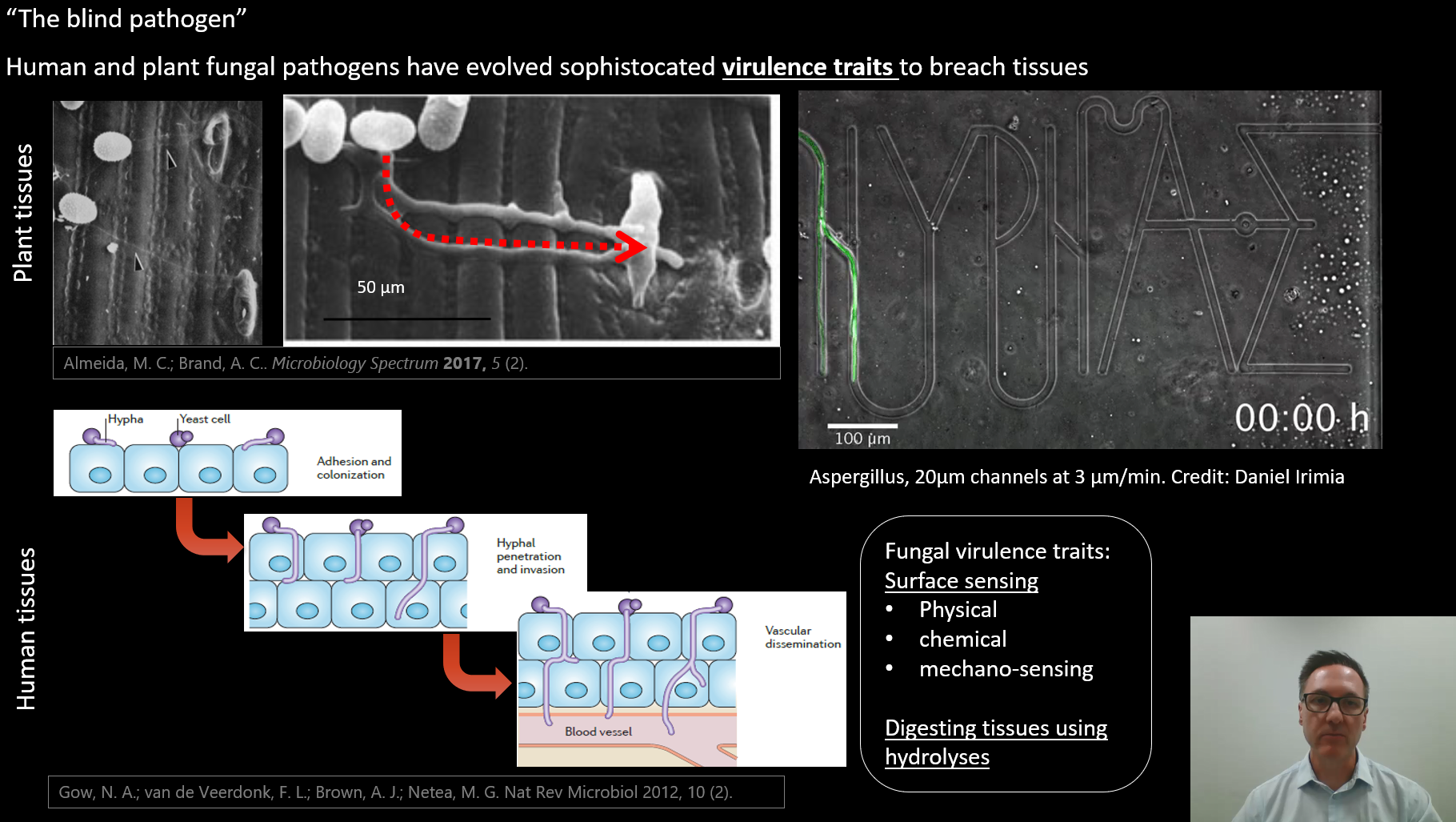
The blind pathogen. Bioinspired and responsive materials for understanding the role of microbial surface sensing in the causes of plant and animal fungal diseases
This post contains information related to my oral presentation at the World Biomaterials Congress 2020.
Questions? Contact?
If you have questions or collaborative ideas, please get in touch.
Email: bryan.coad@adelaide.edu.au
Twitter DM: @DrBryanCoad
Additions & corrections:
On slide 3 I showed the microfluidic device from Prof Daniel Irimia, but failed to give his affiliation. Prof Irimia is researcher and Deputy Director at Massachusetts General Hospital and Harvard Medical School, and organiser of the Fungus Olympics, an event in which my lab participated. You can read more about my involvement in the Fungus Olympics here.
References
Published paper on biodegradable materials:
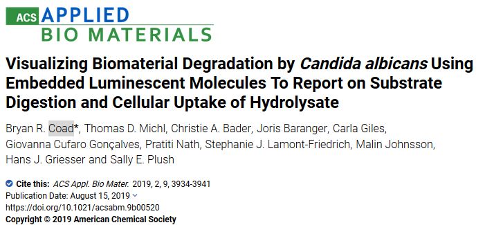
Visualizing Biomaterial Degradation by Candida albicans Using Embedded Luminescent Molecules To Report on Substrate Digestion and Cellular Uptake of Hydrolysate. BR Coad; TD Michl; CA Bader; J Baranger; C Giles; GC Gonçalves; P Nath; SJ Lamont-Friedrich; M Johnsson; HJ Griesser; SE Plush. ACS Applied Bio Materials 2019, 2, 3934-3941. DOI: 10.1021/acsabm.9b00520
Reviews of fungi/plant relationships:
Cell Wall Responses to Biotrophic Fungal Pathogen Invasion. Annual Plant Reviews. J Chowdhury; BR Coad; A Little. 2019, 1001-1030. DOI: 10.1002/9781119312994.apr0634
Perspective on antifungal surface coatings for human health
Anti-infective Surface Coatings: Design and Therapeutic Promise against Device-Associated Infections. BR Coad; HJ Griesser; AY Peleg; A Traven. PLoS Pathogens 2016, 12, e1005598. DOI: 10.1371/journal.ppat.1005598
Information on this website:
Visualizing Biomaterial Degradation by Candida albicans Blog post on this paper
Blog post on the Fungus Olympics
Live cell imaging video of Blumeria graminis on a biomimetic surface coating
Site Navigation
I want to tell the stories behind some of my publications: why the work was done, who funded it, the roles of team members, or stumbling across unexpected results.
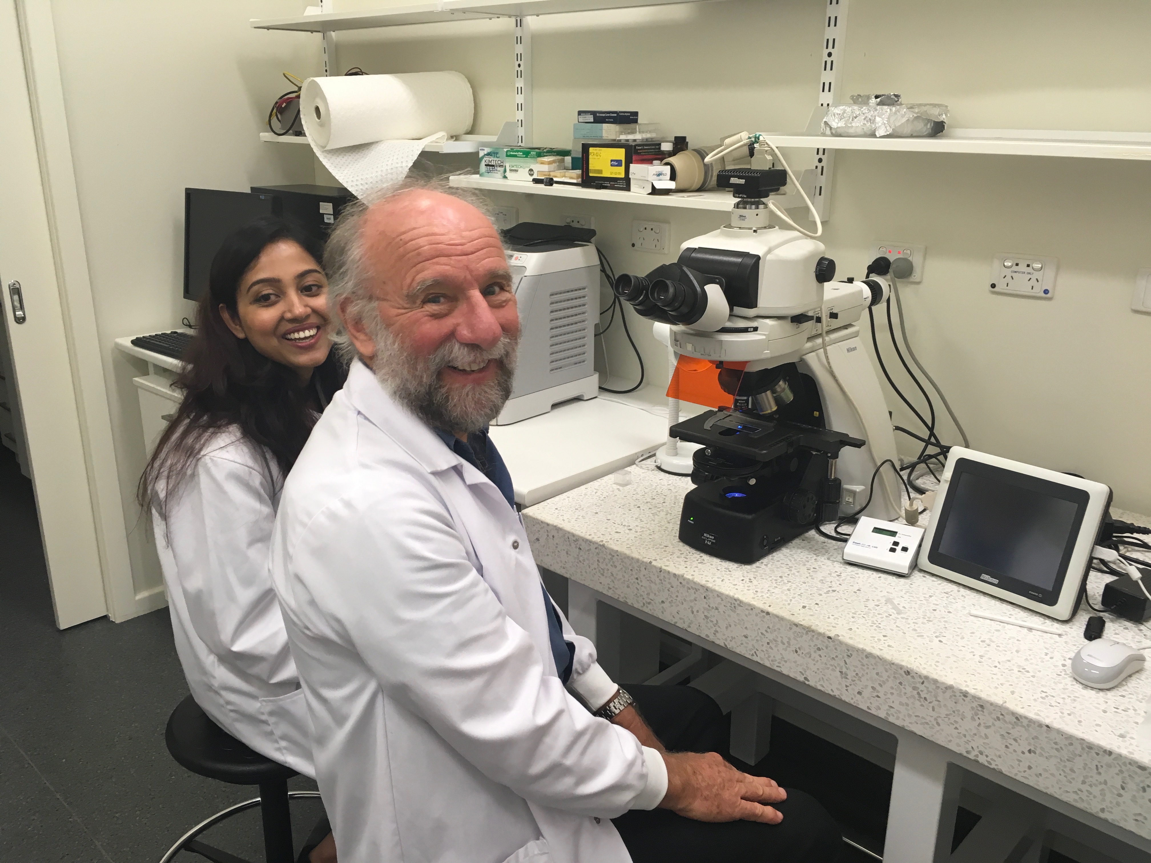
In this post, I’ll tell the story of the team effort behind our latest paper in the journal ACS Applied Bio Materials.
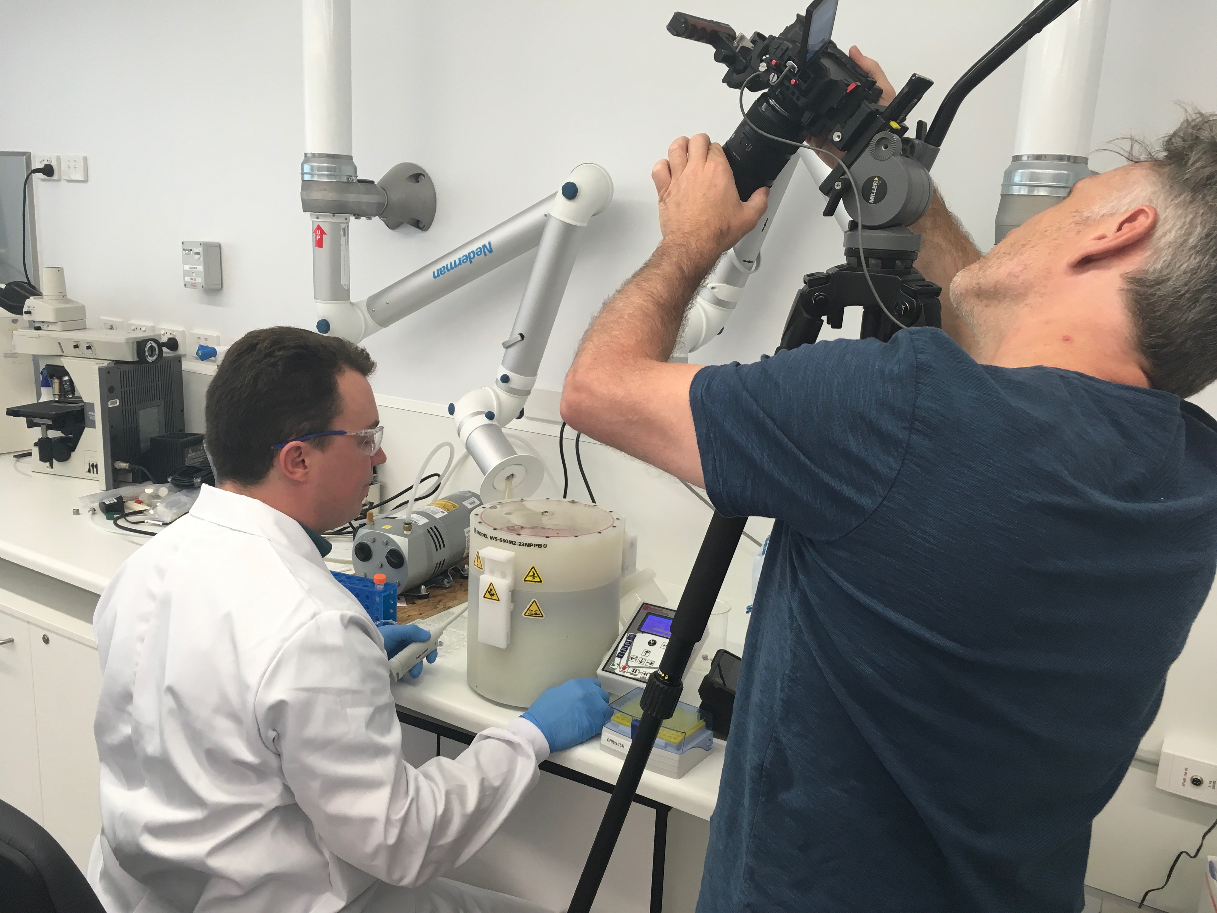
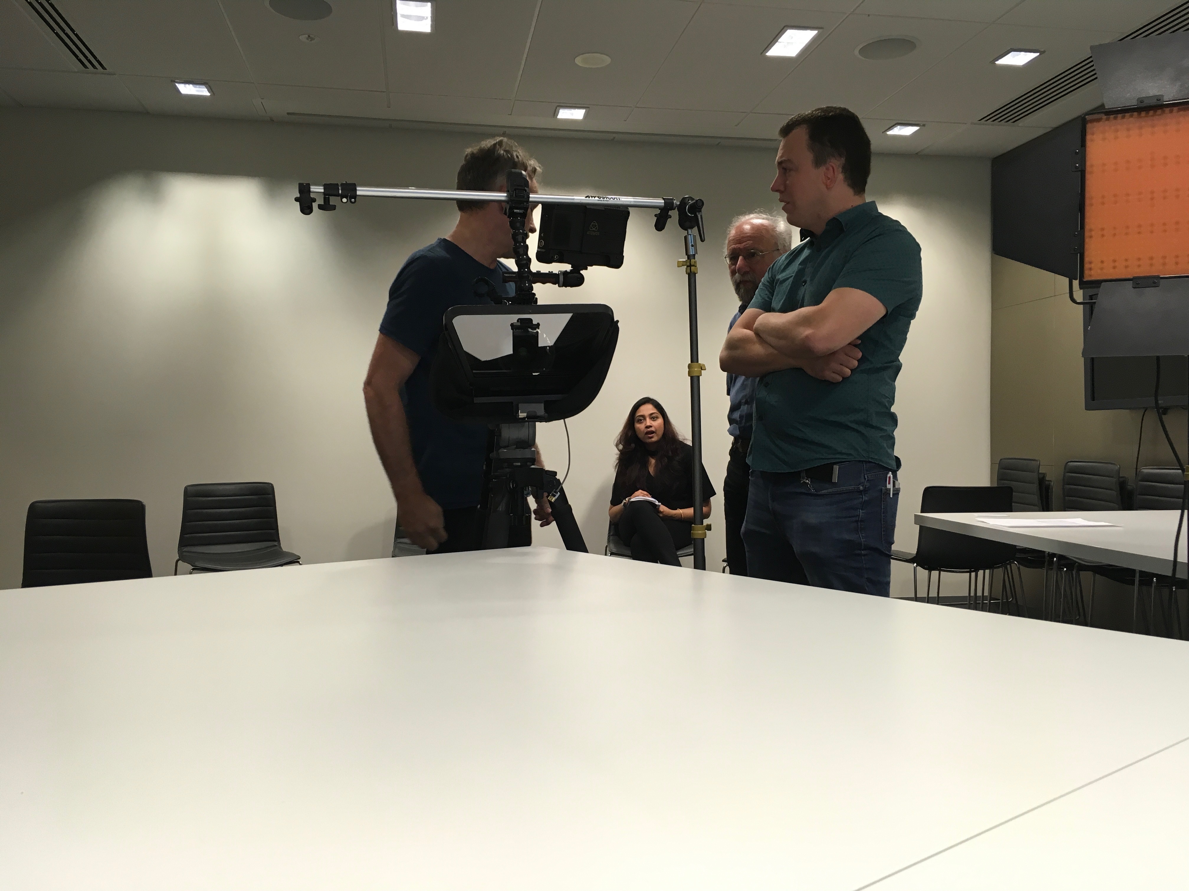
Earlier this year, UniSA produced a video on our Research Themes Investment Scheme project. In this project we describe new materials that release antimicrobial compounds on demand.
Thomas, Hans, Pratiti and I had a lot of fun making this video. Here are some of behind the scenes photos from the production.
The following video shows a time-lapse video of Blumeria graminis f.sp. hordei germination on an artificial surface coating conducted by Dr Bryan Coad and Dr Alan Little, University of Adelaide. The coating was made from hexacosanal (C26 aldehyde) mixed with Formvar ® resin and then cast onto glass slides using the previously published method (1).
Live cell imaging was conducted over 6 hours and compressed into a 5 second video. This video will automatically restart and loop.
In the first hour, primary germ tubes are extended and conidia “tilt” and roll slightly. A small proportion of the conidia develop appressorial germ tubes that extend away from the body. Some of these differentiate into hooked appresoria and attempt penetration of the surface coating. After 6 hours, low humidity in the chamber caused the conidia to desiccate.
This video is used to exemplify the method of live-cell imaging and visualisation of germination on artificial coatings. This technique can be used to visualise germination on other cuticle-mimetic surfaces (such as plasma cuticle mimics).
Using this method, we hope to understand (through visulisation) how fungal germ tubes are developed in response to surface chemistry and topography. Understanding the physicochemical features of surfaces will aid in the discovery of new agents to protect crops from diseases.
References